Molecular biologist. Applied molecular biology
Molecular biology has experienced a period of rapid development of its own research methods, which now differs from biochemistry. These include, in particular, methods of genetic engineering, cloning, artificial expression, and gene knockout. Since DNA is the material carrier of genetic information, molecular biology has become much closer to genetics, and molecular genetics was formed at the junction, which is both a section of genetics and molecular biology. Just as molecular biology makes extensive use of viruses as a research tool, virology uses the methods of molecular biology to solve its problems. Computer technology is involved in the analysis of genetic information, in connection with which new areas of molecular genetics have appeared, which are sometimes considered special disciplines: bioinformatics, genomics and proteomics.
The history of development
This seminal discovery was prepared by a long phase of research into the genetics and biochemistry of viruses and bacteria.
In 1928, Frederick Griffith first showed that an extract of heat-killed pathogenic bacteria could transfer the trait of pathogenicity to benign bacteria. The study of bacterial transformation further led to the purification of the disease agent, which, contrary to expectations, turned out to be not a protein, but a nucleic acid. The nucleic acid itself is not dangerous, it only carries the genes that determine the pathogenicity and other properties of the microorganism.
In the 50s of the XX century, it was shown that bacteria have a primitive sexual process, they are able to exchange extrachromosomal DNA, plasmids. The discovery of plasmids, as well as transformations, formed the basis of the plasmid technology common in molecular biology. Another important discovery for the methodology was the discovery at the beginning of the 20th century of bacterial viruses, bacteriophages. Phages can also transfer genetic material from one bacterial cell to another. Infection of bacteria by phages leads to a change in the composition of bacterial RNA. If, without phages, the composition of RNA is similar to the composition of bacterial DNA, then after infection, RNA becomes more similar to bacteriophage DNA. Thus, it was found that the structure of RNA is determined by the structure of DNA. In turn, the rate of protein synthesis in cells depends on the amount of RNA-protein complexes. This is how it was formulated central dogma of molecular biology: DNA ↔ RNA → protein.
The further development of molecular biology was accompanied by both the development of its methodology, in particular, the invention of a method for determining the nucleotide sequence of DNA (W. Gilbert and F. Sanger, Nobel Prize in Chemistry in 1980), and new discoveries in the field of research into the structure and functioning of genes (see. History of genetics). By the beginning of the 21st century, data were obtained on the primary structure of all human DNA and a number of other organisms, the most important for medicine, agriculture and scientific research, which led to the emergence of several new areas in biology: genomics, bioinformatics, etc.
see also
- Molecular biology (journal)
- Transcriptomics
- Molecular paleontology
- EMBO - European Organization for Molecular Biology
Literature
- Singer M., Berg P. Genes and genomes. - Moscow, 1998.
- Stent G., Kalindar R. Molecular genetics. - Moscow, 1981.
- Sambrook J., Fritsch E.F., Maniatis T. Molecular Cloning. - 1989.
- Patrushev L.I. Expression of genes. - M.: Nauka, 2000. - 000 p., ill. ISBN 5-02-001890-2
Links
Wikimedia Foundation. 2010 .
- Ardatovsky district of the Nizhny Novgorod region
- Arzamas district of the Nizhny Novgorod region
See what "Molecular Biology" is in other dictionaries:
MOLECULAR BIOLOGY- studies the basics. properties and manifestations of life at the molecular level. The most important directions in M. b. are studies of the structural and functional organization of the genetic apparatus of cells and the mechanism for the implementation of hereditary information ... ... Biological encyclopedic dictionary
MOLECULAR BIOLOGY- explores the basic properties and manifestations of life at the molecular level. Finds out how and to what extent the growth and development of organisms, the storage and transmission of hereditary information, the conversion of energy in living cells, and other phenomena are due to ... Big Encyclopedic Dictionary
MOLECULAR BIOLOGY Modern Encyclopedia
MOLECULAR BIOLOGY- MOLECULAR BIOLOGY, the biological study of the structure and function of the MOLECULES that make up living organisms. The main areas of study include the physical and chemical properties of proteins and NUCLEIC ACIDS such as DNA. see also… … Scientific and technical encyclopedic dictionary
molecular biology- a section of biol., which explores the basic properties and manifestations of life at the molecular level. Finds out how and to what extent the growth and development of organisms, the storage and transmission of hereditary information, the conversion of energy in living cells and ... ... Dictionary of microbiology
molecular biology- — Topics of biotechnology EN molecular biology … Technical Translator's Handbook
Molecular biology- MOLECULAR BIOLOGY, explores the basic properties and manifestations of life at the molecular level. Finds out how and to what extent the growth and development of organisms, the storage and transmission of hereditary information, the conversion of energy in living cells and ... ... Illustrated Encyclopedic Dictionary
Molecular biology- a science that sets as its task the knowledge of the nature of life phenomena by studying biological objects and systems at a level approaching the molecular level, and in some cases reaching this limit. The end goal of this is…… Great Soviet Encyclopedia
MOLECULAR BIOLOGY- studies the phenomena of life at the level of macromolecules (ch. arr. proteins and nucleic acids) in cell-free structures (ribosomes, etc.), in viruses, and also in cells. M.'s purpose. establishing the role and mechanism of functioning of these macromolecules based on ... ... Chemical Encyclopedia
molecular biology- explores the basic properties and manifestations of life at the molecular level. Finds out how and to what extent the growth and development of organisms, the storage and transmission of hereditary information, the conversion of energy in living cells and other phenomena ... ... encyclopedic Dictionary
Books
- Molecular biology of the cell. Problem Book, J. Wilson, T. Hunt. The book of American authors is an appendix to the 2nd edition of the textbook `Molecular Biology of the Cell` by B. Alberts, D. Bray, J. Lewis and others. Contains questions and tasks, the purpose of which is to deepen ...
Molecular biology, a science that sets as its task the knowledge of the nature of life phenomena by studying biological objects and systems at a level approaching the molecular level, and in some cases reaching this limit. The ultimate goal in this case is to clarify how and to what extent the characteristic manifestations of life, such as heredity, reproduction of one's own kind, protein biosynthesis, excitability, growth and development, storage and transmission of information, energy transformations, mobility, etc. , are due to the structure, properties and interaction of molecules of biologically important substances, primarily the two main classes of high-molecular biopolymers - proteins and nucleic acids. A distinctive feature of M. b. - the study of the phenomena of life on inanimate objects or those that are characterized by the most primitive manifestations of life. These are biological formations from the cellular level and below: subcellular organelles, such as isolated cell nuclei, mitochondria, ribosomes, chromosomes, cell membranes; further - systems standing on the border of animate and inanimate nature - viruses, including bacteriophages, and ending with the molecules of the most important components of living matter - nucleic acids and proteins.
The foundation on which M. developed. was laid by such sciences as genetics, biochemistry, physiology of elementary processes, etc. According to the origins of its development, M. b. is inextricably linked to molecular genetics, which continues to be an important part of
A distinctive feature of M. b. is its three-dimensionality. The essence of M. b. M. Perutz sees it in interpreting biological functions in terms of molecular structure. M. b. aims to get answers to the question "how", knowing the essence of the role and participation of the entire structure of the molecule, and to the questions "why" and "why", having found out, on the one hand, the relationship between the properties of the molecule (again, primarily proteins and nucleic acids) and the functions it performs and, on the other hand, the role of such individual functions in the overall complex of manifestations of vital activity.
The most important achievements of molecular biology. Here is a far from complete list of these achievements: disclosure of the structure and mechanism of the biological function of DNA, all types of RNA and ribosomes, disclosure of the genetic code; discovery of reverse transcription, i.e., DNA synthesis on an RNA template; study of the mechanisms of functioning of respiratory pigments; discovery of the three-dimensional structure and its functional role in the action of enzymes, the principle of matrix synthesis and the mechanisms of protein biosynthesis; disclosure of the structure of viruses and the mechanisms of their replication, the primary and, in part, the spatial structure of antibodies; isolation of individual genes, chemical and then biological (enzymatic) gene synthesis, including human, outside the cell (in vitro); transfer of genes from one organism to another, including into human cells; the rapidly progressing deciphering of the chemical structure of an increasing number of individual proteins, mainly enzymes, as well as nucleic acids; discovery of the phenomena of "self-assembly" of some biological objects of ever-increasing complexity, starting from nucleic acid molecules and moving on to multicomponent enzymes, viruses, ribosomes, etc.; elucidation of allosteric and other basic principles of regulation of biological functions and processes.
Problems of molecular biology. Along with the specified important tasks M. would. (knowledge of the laws of "recognition", self-assembly and integration) the actual direction of scientific search for the near future is the development of methods that allow deciphering the structure, and then the three-dimensional, spatial organization of high-molecular nucleic acids. All the most important methods, the use of which ensured the emergence and success of M. b., were proposed and developed by physicists (ultracentrifugation, X-ray diffraction analysis, electron microscopy, nuclear magnetic resonance, etc.). Almost all new physical experimental approaches (for example, the use of computers, synchrotron, or bremsstrahlung, radiation, laser technology, and others) open up new possibilities for an in-depth study of the problems of M. b. Among the most important tasks of a practical nature, the answer to which is expected from M. b., in the first place is the problem of the molecular basis of malignant growth, then - ways to prevent, and perhaps overcome hereditary diseases - "molecular diseases". Of great importance will be the elucidation of the molecular basis of biological catalysis, ie, the action of enzymes. Among the most important modern directions of M. b. should include the desire to decipher the molecular mechanisms of action of hormones, toxic and medicinal substances, as well as to find out the details of the molecular structure and functioning of such cellular structures as biological membranes involved in the regulation of the processes of penetration and transport of substances. More distant goals M. b. - knowledge of the nature of nervous processes, mechanisms of memory, etc. One of the important emerging sections of M. b. - so-called. genetic engineering, which sets as its task the purposeful operation of the genetic apparatus (genome) of living organisms, starting with microbes and lower (single-celled) and ending with humans (in the latter case, primarily for the purpose of radical treatment of hereditary diseases and correction of genetic defects).
The most important directions of the MB:
- Molecular genetics - the study of the structural and functional organization of the genetic apparatus of the cell and the mechanism for the implementation of hereditary information
– Molecular virology – the study of the molecular mechanisms of the interaction of viruses with cells
– Molecular immunology – the study of patterns of immune reactions of the body
– Molecular biology of development – the study of the appearance of cell diversity in the course of individual development of organisms and specialization of cells
Main objects of research: Viruses (including bacteriophages), Cells and subcellular structures, Macromolecules, Multicellular organisms.
The development of biochemistry, biophysics, genetics, cytochemistry, many sections of microbiology and virology around the beginning of the 40s of the XX century. closely led to the study of life phenomena at the molecular level. The successes achieved by these sciences, simultaneously and from different sides, led to the realization of the fact that it is at the molecular level that the main control systems of the body function and that the further progress of these sciences will depend on the disclosure of the biological functions of the molecules that make up the bodies of organisms, their participation in the synthesis and disintegration, mutual transformations and reproduction of compounds in the cell, as well as the exchange of energy and information that occurs in this case. Thus, at the junction of these biological disciplines with chemistry and physics, a completely new branch arose - molecular biology.
Unlike biochemistry, the attention of modern molecular biology is focused mainly on the study of the structure and function of the most important classes of biopolymers - proteins and nucleic acids, the first of which determine the very possibility of metabolic reactions, and the second - the biosynthesis of specific proteins. It is clear, therefore, that it is impossible to make a clear distinction between molecular biology and biochemistry, the corresponding branches of genetics, microbiology, and virology.
The emergence of molecular biology was closely associated with the development of new research methods, which have already been discussed in the relevant chapters. Along with the development of electron microscopy and other methods of microscopic technique, the methods of fractionation of cellular elements developed in the 1950s played an important role. They were based on improved methods of differential centrifugation (A. Claude, 1954). By this time, there were already quite reliable methods for the isolation and fractionation of biopolymers. This includes, in particular, the method of protein fractionation by electrophoresis proposed by A. Tiselius (1937; Nobel Prize, 1948), methods for isolating and purifying nucleic acids (E. Kay, A. Downs, M. Sevag, A. Mirsky, and others. ). At the same time, various methods of chromatographic analysis were developed in many laboratories of the world (A. Martin and R. Sing, 1941; Nobel Prize, 1952), subsequently significantly improved.
X-ray diffraction analysis played an invaluable service in deciphering the structure of biopolymers. The basic principles of X-ray diffraction analysis were developed at King's College London University under the leadership of W. Bragg by a group of researchers, which included J. Bernal, A. Londsdale, W. Astbury, J. Robertson and others.
Special mention should be made of the studies of Protoplasm Biochemistry (1925 - 1929), Professor of Moscow State University A. R. Kizel, which were of great importance for the subsequent development of molecular biology. Kizel dealt a blow to the firmly rooted notion that any protoplasm is based on a special protein body - plates, which allegedly determines all its most important structural and functional features. He showed that plates are a protein that is found only in myxomycetes, and then at a certain stage of development, and that no permanent component - a single skeletal protein - exists in protoplasm. Thus, the study of the problem of the structure of protoplasm and the functional role of proteins took the right path and received scope for its development. Kisel's research has won worldwide recognition, stimulating the study of the chemistry of the constituent parts of the cell.
The term "molecular biology", first used by the English crystallographer Professor of the University of Leeds W. Astbury, probably appeared in the early 1940s (before 1945). The fundamental X-ray diffraction studies of proteins and DNA, carried out by Astbury in the 1930s, served as the basis for the subsequent successful deciphering of the secondary structure of these biopolymers. In 1963, J. Bernal wrote: "A monument to him will be erected by the whole of molecular biology - the science that he named and really founded" * , In the literature, this term appeared for the first time, perhaps, in 1946 in the article by W. Astbury "Progress of X-ray diffraction analysis of organic and fibrillar compounds", published in the English journal "Nature" ** . In his Harvey Lecture, Astbury (1950) noted: “I am pleased that the term molecular biology is now quite widely used, although it is unlikely that I was the first to propose it. I liked it and I have long tried to spread it” ***. Already in 1950 Astbury was clear that molecular biology deals primarily with the structure and conformation of macromolecules, the study of which is of decisive importance for understanding the functioning of living organisms.
* (biogr. Mem. Fellows Roy. Soc, 1963, v. 9, 29.)
** (W. T. Astbury. Progress of X-ray analysis of organic and fiber structures.- Nature,. 1946, v. 157, 121.)
*** (W. T. Astbury. Adventures in Molecular Biology. Thomas Springfield, 1952, p. 3.)
Molecular biology has faced and faces, in fact, the same tasks as biology as a whole - the knowledge of the essence of life and its basic phenomena, in particular, such as heredity and variability. Modern molecular biology is primarily intended to decipher the structure and function of genes, the ways and mechanisms of realization of the genetic information of organisms at different stages of ontogenesis and at different stages of its reading. It is designed to reveal the subtle mechanisms of regulation of gene activity and cell differentiation, to elucidate the nature of mutagenesis and the molecular basis of the evolutionary process.
Establishing the genetic role of nucleic acids
For the development of molecular biology, the following discoveries were of the greatest importance. In 1944, American researchers O. Avery, K. McLeod (Nobel Prize, 1923) and M. McCarthy showed that DNA molecules isolated from pneumococci have transforming activity. After hydrolysis of these DNAs by deoxyribonuclease, their transforming activity completely disappeared. Thus, for the first time, it was convincingly proved that it is DNA, and not protein, that is endowed with genetic functions in a cell.
In fairness, it should be noted that the phenomenon of bacterial transformation was discovered much earlier than the discovery of Avery, McLeod and McCarthy. In 1928, F. Griffith published an article in which he reported that after adding killed cells of an encapsulated virulent strain to non-virulent (non-encapsulated) pneumococci, the resulting mixture of cells becomes fatal for mice. Moreover, live pneumococcal cells isolated from animals infected with this mixture were already virulent and possessed a polysaccharide capsule. Thus, in this experiment, it was shown that under the influence of some components of the killed pneumococcal cells, the non-encapsulated form of bacteria turns into a capsule-forming virulent form. Sixteen years later, Avery, McLeod, and McCarthy replaced killed whole pneumococcal cells with their deoxyribonucleic acid in this experiment and showed that it was DNA that had transforming activity (see also chapters 7 and 25). The significance of this discovery is difficult to overestimate. It stimulated the study of nucleic acids in many laboratories around the world and forced scientists to focus on DNA.
Along with the discovery of Avery, McLeod, and McCarthy, by the beginning of the 1950s, a fairly large amount of direct and indirect evidence had already accumulated that nucleic acids play an exceptional role in life and carry a genetic function. This, in particular, was indicated by the nature of DNA localization in the cell and the data of R. Vendrelli (1948) that the DNA content per cell is strictly constant and correlates with the degree of ploidy: in haploid germ cells, DNA is half that in diploid somatic cells. The pronounced metabolic stability of DNA also testified in favor of the genetic role of DNA. By the beginning of the 1950s, a lot of various facts had accumulated, indicating that most of the known mutagenic factors act mainly on nucleic acids and, in particular, on DNA (R. Hotchkiss, 1949; G. Ephrussi-Taylor, 1951; E. Freese , 1957 and others).
Of particular importance in establishing the genetic role of nucleic acids was the study of various phages and viruses. In 1933, D. Schlesinger found DNA in the bacteriophage of Escherichia coli. Since the isolation of tobacco mosaic virus (TMV) in the crystalline state by W. Stanley (1935, Nobel Prize, 1946), a new stage in the study of plant viruses has begun. In 1937 - 1938. employees of the Rothamsted Agricultural Station (England) F. Bowden and N. Peary showed that many plant viruses isolated by them are not globulins, but are ribonucleoproteins and contain nucleic acid as an obligatory component. At the very beginning of the 40s, the works of G. Schramm (1940), P. A. Agatov (1941), G. Miller and W. Stanley (1941) were published, indicating that a noticeable chemical modification of the protein component does not lead to loss of TMV infectivity. This indicated that the protein component could not be the carrier of the hereditary properties of the virus, as many microbiologists continued to believe. Convincing evidence in favor of the genetic role of nucleic acid (RNA) in plant viruses was obtained in 1956 by G. Schramm in Tübingen (FRG) and H. Frenkel-Konrath in California (USA). These researchers almost simultaneously and independently of each other isolated RNA from TMV and showed that it, and not protein, has infectivity: as a result of infection of tobacco plants with this RNA, normal viral particles were formed and multiplied in them. This meant that RNA contained information for the synthesis and assembly of all viral components, including the viral protein. In 1968, I. G. Atabekov established that protein plays a significant role in the very infection of plants - the nature of the protein determines the spectrum of host plants.
In 1957, Frenkel-Konrat for the first time carried out the reconstruction of the TMV from its constituent components - RNA and protein. Along with normal particles, he received mixed "hybrids" in which the RNA was from one strain and the protein from another. The heredity of such hybrids was completely determined by RNA, and the progeny of the viruses belonged to the strain whose RNA was used to obtain the initial mixed particles. Later, the experiments of A. Gierer, G. Schuster and G. Schramm (1958) and G. Witman (1960 - 1966) showed that the chemical modification of the TMV nucleic component leads to the appearance of various mutants of this virus.
In 1970, D. Baltimore and G. Temin found that the transfer of genetic information can occur not only from DNA to RNA, but vice versa. They found in some oncogenic RNA-containing viruses (oncornaviruses) a special enzyme, the so-called reverse transcriptase, which is capable of synthesizing complementary DNA on RNA chains. This major discovery made it possible to understand the mechanism of insertion of the genetic information of RNA-containing viruses into the host genome and to take a fresh look at the nature of their oncogenic action.
Discovery of nucleic acids and study of their properties
The term nucleic acids was introduced by the German biochemist R. Altman in 1889, after these compounds were discovered in 1869 by the Swiss physician F. Miescher. Misher extracted the pus cells with dilute hydrochloric acid for several weeks and obtained almost pure nuclear material in the remainder. He considered this material to be a characteristic "substance of cell nuclei and called it nuclein. In its properties, nuclein differed sharply from proteins: it was more acidic, did not contain sulfur, but it contained a lot of phosphorus, it was readily soluble in alkalis, but did not dissolve in dilute acids.
Misher sent the results of his observations on nuclein to F. Goppe-Seyler for publication in a journal. The substance he described was so unusual (at that time only lecithin was known of all biological phosphorus-containing compounds) that Goppe-Seyler did not believe Misher's experiments, returned the manuscript to him and instructed his employees N. Plosh and N. Lyubavin to check his conclusions on other material . Miescher's work "On the chemical composition of pus cells" was published two years later (1871). At the same time, the works of Goppe-Seyler and his collaborators were published on the composition of pus cells, erythrocytes of birds, snakes, and other cells. Over the next three years, nuclein was isolated from animal cells and yeast.
In his work, Misher noted that a detailed study of different nucleins can lead to the establishment of differences between them, thereby anticipating the idea of specificity of nucleic acids. While studying salmon milk, Misher found that the nuclein in them is in the form of salt and is associated with the main protein, which he called protamine.
In 1879, A. Kossel began to study nucleins in the laboratory of Goppe-Seyler. In 1881, he isolated hypoxanthine from nuclein, but at that time he still doubted the origin of this base and believed that hypoxanthine could be a degradation product of proteins. In 1891, among the products of nuclein hydrolysis, Kossel discovered adenine, guanine, phosphoric acid, and another substance with the properties of sugar. For research on the chemistry of nucleic acids, Kossel was awarded the Nobel Prize in 1910.
Further progress in deciphering the structure of nucleic acids is associated with the research of P. Levin and colleagues (1911 - 1934). In 1911, P. Levin and V. Jacobs identified the carbohydrate component of adenosine and guanosine; they found that these nucleosides contain D-ribose. In 1930, Lewin showed that the carbohydrate component of deoxyribonucleosides is 2-deoxy-D-ribose. From his work, it became known that nucleic acids are built from nucleotides, i.e., phosphorylated nucleosides. Levin believed that the main type of bond in nucleic acids (RNA) is the 2", 5" phosphodiester bond. This notion turned out to be wrong. Thanks to the work of the English chemist A. Todd (Nobel Prize, 1957) and his collaborators, as well as the English biochemists R. Markham and J. Smith, it became known in the early 50s that the main type of bond in RNA is 3", 5" - phosphodiester bond.
Lewin showed that different nucleic acids can differ in the nature of the carbohydrate component: some of them contain the sugar deoxyribose, while others contain ribose. In addition, these two types of nucleic acids differed in the nature of one of the bases: pentose-type nucleic acids contained uracil, and deoxypentose-type nucleic acids contained thymine. Deoxypentose nucleic acid (in modern terminology, deoxyribonucleic acid - DNA) was usually easily isolated in large quantities from the thymus (sweet gland) of calves. Therefore, it was called thymonucleic acid. The source of pentose-type nucleic acid (RNA) was mainly yeast and wheat germ. This type was often referred to as yeast nucleic acid.
In the early 1930s, the notion that plant cells were characterized by a yeast-type nucleic acid was rather firmly rooted, while thymonucleic acid was characteristic only of the nuclei of animal cells. The two types of nucleic acids, RNA and DNA, were then called plant and animal nucleic acids, respectively. However, as the early studies of A. N. Belozersky showed, such a division of nucleic acids is unjustified. In 1934, Belozersky first discovered thymonucleic acid in plant cells: from pea seedlings, he isolated and identified the thymine-pyrimidine base, which is characteristic of DNA. Then he discovered thymine in other plants (soybean seeds, beans). In 1936, A. N. Belozersky and I. I. Dubrovskaya isolated DNA preparatively from horse chestnut seedlings. In addition, a series of studies carried out in England in the 1940s by D. Davidson and co-workers convincingly showed that plant nucleic acid (RNA) is contained in many animal cells.
The widespread use of the cytochemical reaction for DNA developed by R. Felgen and G. Rosenbeck (1924) and the reaction of J. Brachet (1944) for RNA made it possible to quickly and unambiguously resolve the issue of the preferential localization of these nucleic acids in the cell. It turned out that DNA is concentrated in the nucleus, while RNA is predominantly concentrated in the cytoplasm. Later, it was found that RNA is contained both in the cytoplasm and in the nucleus, and in addition, cytoplasmic DNA was identified.
As for the question of the primary structure of nucleic acids, by the mid-1940s, P. Levin's idea was firmly established in science, according to which all nucleic acids are built according to the same type and consist of the same so-called tetranucleotide blocks. Each of these blocks, according to Lewin, contains four different nucleotides. The tetranucleotide theory of the structure of nucleic acids largely deprived these biopolymers of specificity. Therefore, it is not surprising that at that time all the specifics of living things were associated only with proteins, the nature of the monomers of which is much more diverse (20 amino acids).
The first gap in the theory of the tetranucleotide structure of nucleic acids was made by the analytical data of the English chemist J. Gouland (1945 - 1947). When determining the composition of nucleic acids by the base nitrogen, he did not obtain an equimolar ratio of bases, as it should have been according to Lewin's theory. Finally, the tetranucleotide theory of the structure of nucleic acids collapsed as a result of the research of E. Chargaff and his collaborators (1949 - 1951). Chargaff used paper chromatography to separate the bases released from DNA as a result of its acid hydrolysis. Each of these bases was accurately determined spectrophotometrically. Chargaff noticed significant deviations from the equimolar ratio of bases in DNA of different origins and for the first time definitely stated that DNA has a pronounced species specificity. This ended the hegemony of the concept of protein specificity in the living cell. Analyzing DNA of different origins, Chargaff discovered and formulated unique patterns of DNA composition, which entered science under the name of Chargaff's rules. According to these rules, in all DNA, regardless of origin, the amount of adenine is equal to the amount of thymine (A = T), the amount of guanine is equal to the amount of cytosine (G = C), the amount of purines is equal to the amount of pyrimidines (G + A = C + T), the amount bases with 6-amino groups is equal to the number of bases with 6-keto groups (A + C = G + T). At the same time, despite such strict quantitative correspondences, DNA of different species differ in the value of the A+T:G+C ratio. In some DNA, the amount of guanine and cytosine prevails over the amount of adenine and thymine (Chargaff called these DNA GC-type DNA); other DNAs contained more adenine and thymine than guanine and cytosine (these DNAs were called AT-type DNA). The data obtained by Chargaff on the composition of DNA played an exceptional role in molecular biology. It was they that formed the basis for the discovery of the structure of DNA, made in 1953 by J. Watson and F. Crick.
Back in 1938, W. Astbury and F. Bell, using X-ray diffraction analysis, showed that the base planes in DNA should be perpendicular to the long axis of the molecule and resemble, as it were, a stack of plates lying on top of each other. With the improvement of the technique of X-ray diffraction analysis, by 1952 - 1953. accumulated information that made it possible to judge the length of individual bonds and the angles of inclination. This made it possible to represent with the greatest probability the nature of the orientation of the rings of pentose residues in the sugar-phosphate backbone of the DNA molecule. In 1952, S. Farberg proposed two speculative models of DNA, which represented a single-stranded molecule folded or twisted on itself. A no less speculative model of the structure of DNA was proposed in 1953 by L. Pauling (Nobel Prize winner, 1954) and R. Corey. In this model, three twisted strands of DNA formed a long helix, the core of which was represented by phosphate groups, and the bases were located outside of it. By 1953, M. Wilkins and R. Franklin obtained clearer X-ray diffraction patterns of DNA. Their analysis showed the complete failure of the models of Farberg, Pauling and Corey. Using Chargaff's data, comparing different combinations of molecular models of individual monomers and X-ray diffraction data, J. Watson and F. Crick in 1953 came to the conclusion that the DNA molecule must be a double-stranded helix. Chargaff's rules severely limited the number of possible ordered combinations of bases in the proposed DNA model; they suggested to Watson and Crick that there must be a specific base pairing in the DNA molecule - adenine with thymine, and guanine with cytosine. In other words, adenine in one strand of DNA always strictly corresponds to thymine in the other strand, and guanine in one strand necessarily corresponds to cytosine in the other. Thus, Watson and Crick were the first to formulate the extremely important principle of the complementary structure of DNA, according to which one DNA strand complements another, i.e., the base sequence of one strand uniquely determines the base sequence in the other (complementary) strand. It became obvious that already in the very structure of DNA lies the potential for its exact reproduction. This model of DNA structure is currently generally accepted. Crick, Watson and Wilkins were awarded the Nobel Prize in 1962 for deciphering the structure of DNA.
It should be noted that the idea of a mechanism for the exact reproduction of macromolecules and the transmission of hereditary information originated in our country. In 1927, N. K. Koltsov suggested that during cell reproduction, the reproduction of molecules occurs by exact autocatalytic reproduction of the existing parent molecules. True, at that time Koltsov endowed this property not with DNA molecules, but with molecules of a protein nature, the functional significance of which was then unknown. Nevertheless, the very idea of autocatalytic reproduction of macromolecules and the mechanism of transmission of hereditary properties turned out to be prophetic: it became the guiding idea of modern molecular biology.
Conducted in the laboratory of A. N. Belozersky by A. S. Spirin, G. N. Zaitseva, B. F. Vanyushin, S. O. Uryson, A. S. Antonov and others variety of organisms fully confirmed the patterns discovered by Chargaff, and full compliance with the molecular model of the structure of DNA proposed by Watson and Crick. These studies have shown that the DNA of different bacteria, fungi, algae, actinomycetes, higher plants, invertebrates and vertebrates have a specific composition. Differences in the composition (the content of AT-base pairs) are especially pronounced in microorganisms, turning out to be an important taxonomic feature. In higher plants and animals, species variations in the composition of DNA are much less pronounced. But this does not mean that their DNA is less specific. In addition to the composition of bases, specificity is largely determined by their sequence in DNA chains.
Along with the usual bases, additional nitrogenous bases were found in DNA and RNA. Thus, G. White (1950) found 5-methylcytosine in the DNA of plants and animals, and D. Dunn and J. Smith (1958) found methylated adenine in some DNA. For a long time, methylcytosine was considered a hallmark of the genetic material of higher organisms. In 1968, A. N. Belozersky, B. F. Vanyushin and N. A. Kokurina found that it can also be found in the DNA of bacteria.
In 1964, M. Gold and J. Hurwitz discovered a new class of enzymes that carry out the natural modification of DNA - its methylation. After this discovery, it became clear that minor (contained in small amounts) bases arise already on the finished DNA polynucleotide chain as a result of specific methylation of cytosine and adenine residues in special sequences. In particular, according to B. F. Vanyushin, Ya. I. Buryanov, and A. N. Belozersky (1969), adenine methylation in E. coli DNA can occur in terminating codons. According to A. N. Belozersky and colleagues (1968 - 1970), as well as M. Meselson (USA) and V. Arber (Switzerland) (1965 - 1969), methylation gives unique individual features to DNA molecules and, in combination with the action of specific nucleases, is part of a complex mechanism that controls the synthesis of DNA in the cell. In other words, the nature of methylation of a particular DNA predetermines the question of whether it can multiply in a given cell.
Almost at the same time, the isolation and intensive study of DNA methylases and restriction endonucleases began; in 1969 - 1975 the nucleotide sequences recognized in DNA by some of these enzymes have been established (X. Boyer, X. Smith, S. Lynn, K. Murray). When different DNAs are hydrolyzed by a restriction enzyme, rather large fragments with identical "sticky" ends are cleaved out. This makes it possible not only to analyze the structure of genes, as is done in small viruses (D. Nathans, S. Adler, 1973 - 1975), but also to construct various genomes. With the discovery of these specific restriction enzymes, genetic engineering has become a tangible reality. Embedded in small plasmid DNA genes of various origins are already easily introduced into various cells. So, a new type of biologically active plasmids was obtained, giving resistance to certain antibiotics (S. Cohen, 1973), ribosomal genes of a frog and Drosophila were introduced into Escherichia coli plasmids (J. Morrow, 1974; X. Boyer, D. Hogness, R. Davis , 1974 - 1975). Thus, real ways are open for obtaining fundamentally new organisms by introducing and integrating various genes into their gene pool. This discovery can be directed to the benefit of all mankind.
In 1952, G. White and S. Cohen discovered that the DNA of T-even phages contains an unusual base - 5-hydroxymethylcytosine. Later, from the works of E. Volkin and R. Sinsheimer (1954) and Cohen (1956), it became known that hydroxymethylcytosine residues can be completely or partially glucosidized, as a result of which the phage DNA molecule is protected from the hydrolytic action of nucleases.
In the early 1950s, from the works of D. Dunn and J. Smith (England), S. Zamenhof (USA) and A. Wacker (Germany), it became known that many artificial base analogues can be included in DNA, sometimes replacing up to 50% thymine. As a rule, these substitutions lead to errors in DNA replication, transcription and translation and to the appearance of mutants. Thus, J. Marmur (1962) found that the DNA of some phages contains oxymethyluracil instead of thymine. In 1963, I. Takahashi and J. Marmur discovered that the DNA of one of the phages contains uracil instead of thymine. Thus, another principle, according to which nucleic acids were previously separated, collapsed. Since the time of P. Levin's work, it has been believed that thymine is the hallmark of DNA, and uracil is the hallmark of RNA. It became clear that this sign is not always reliable, and the fundamental difference in the chemical nature of the two types of nucleic acids, as it seems today, is only the nature of the carbohydrate component.
In the study of phages, many unusual features of the organization of nucleic acids have been uncovered. Since 1953, it has been believed that all DNA are double-stranded linear molecules, while RNA is only single-stranded. This position was significantly shaken in 1961, when R. Sinsheimer discovered that the DNA of the phage φ X 174 is represented by a single-stranded circular molecule. However, later it turned out that in this form this DNA exists only in a vegetative phage particle, and the replicative form of the DNA of this phage is also double-stranded. In addition, it turned out to be quite unexpected that the RNA of some viruses can be double-stranded. This new type of macromolecular organization of RNA was discovered in 1962 by P. Gomatos, I. Tamm and other researchers in some animal viruses and in plant wound tumor virus. Recently, V. I. Agol and A. A. Bogdanov (1970) established that in addition to linear RNA molecules, there are also closed or cyclic molecules. They detected cyclic double-stranded RNA, in particular, in the encephalomyelocarditis virus. Thanks to the works of X. Deveaux, L. Tinoko, T. I. Tikhonenko, E. I. Budovsky and others (1960 - 1974), the main features of the organization (laying) of genetic material in bacteriophages became known.
In the late 1950s, the American scientist P. Doty found that heating causes DNA denaturation, which is accompanied by the breaking of hydrogen bonds between base pairs and the separation of complementary chains. This process has the character of a "spiral-coil" phase transition and resembles the melting of crystals. Therefore, Doty called the process of thermal denaturation of DNA DNA melting. With slow cooling, renaturation of molecules occurs, i.e., the reunification of complementary halves.
The principle of renaturation in 1960 was used by J. Marmur and K. Schildkraut to determine the degree of "hybridizability" of DNA of different microorganisms. Subsequently, E. Bolton and B. McCarthy improved this technique by proposing the method of the so-called DNA-agar columns. This method turned out to be indispensable in studying the degree of homology of the nucleotide sequence of different DNA and elucidating the genetic relationship of different organisms. The denaturation of DNA discovered by Doty in combination with the chromatography on methylated albumin described by J. Mandel and A. Hershey * (1960) and centrifugation in a density gradient (the method was developed in 1957 by M. Meselson, F. Stahl and D. Winograd) is widely used for separation, isolation and analysis of individual complementary DNA strands For example, W. Shibalsky (USA), using these techniques to separate the DNA of the lambda phage, showed in 1967 - 1969 that both phage chains are genetically active, and not one, as this was considered to be (S. Spiegelman, 1961). It should be noted that for the first time the idea of the genetic significance of both DNA strands of the lambda phage was expressed in the USSR by SE Bresler (1961).
* (For their work on the genetics of bacteria and viruses, A. Hershey, together with M. Delbrück and S. Luria, were awarded the Nobel Prize in 1969.)
To understand the organization and functional activity of the genome, the determination of the DNA nucleotide sequence is of paramount importance. The search for methods for such determination is carried out in many laboratories around the world. Since the late 1950s, M. Beer and his collaborators have been trying to establish the DNA sequence using electron microscopy in the USA, but so far without success. In the early 1950s, from the first works of Sinsheimer, Chargaff, and other researchers on the enzymatic degradation of DNA, it became known that different nucleotides in a DNA molecule are distributed, although not randomly, but unevenly. According to the English chemist C. Barton (1961), pyrimidines (more than 70%) are concentrated mainly in the form of the corresponding blocks. A. L. Mazin and B. F. Vanyushin (1968 - 1969) found that different DNAs have different degrees of pyrimidine cohesion and that in the DNA of animal organisms it increases markedly as it moves from lower to higher. Thus, the evolution of organisms is also reflected in the structure of their genomes. That is why, for understanding the evolutionary process as a whole, the comparative study of the structure of nucleic acids is of particular importance. Analysis of the structure of biologically important polymers and, first of all, DNA is extremely important for solving many particular problems of phylogenetics and taxonomy.
It is interesting to note that the English physiologist E. Lankester, who studied the hemoglobins of mollusks, anticipated the ideas of molecular biology exactly 100 years ago, wrote: “Chemical differences between different species and genera of animals and plants are as important for clarifying the history of their origin as their form. If we could clearly establish the differences in the molecular organization and functioning of organisms, we would be able to understand the origin and evolution of different organisms much better than on the basis of morphological observations " * . The significance of biochemical studies for taxonomy was also emphasized by VL Komarov, who wrote that "the basis of all even purely morphological features, on the basis of which we classify and establish species, are precisely biochemical differences" ** .
* (E. R. Lankester. Uber das Vorcommen von Haemoglobin in den Muskeln der Mollusken und die Verbreitung desselben in den lebendigen Organismen.- "Pfluger" s Archiv fur die gesammte Physiol., 1871, Bd 4, 319.)
** (V. L. Komarov. Selected works, vol. 1. M.-L., Publishing House of the Academy of Sciences of the USSR, 1945, p. 331.)
A. V. Blagoveshchenskii and S. L. Ivanov, back in the 1920s, took the first steps in our country to elucidate certain questions of the evolution and systematics of organisms on the basis of a comparative analysis of their biochemical composition (see Chapter 2). Comparative analysis of the structure of proteins and nucleic acids is now becoming an increasingly tangible tool for taxonomists (see Chapter 21). This method of molecular biology allows not only to clarify the position of individual species in the system, but also makes it necessary to take a fresh look at the very principles of classification of organisms, and sometimes to revise the entire system as a whole, as happened, for example, with the systematics of microorganisms. Undoubtedly, in the future, the analysis of the structure of the genome will occupy a central place in the chemosystematics of organisms.
Of great importance for the development of molecular biology was the deciphering of the mechanisms of DNA replication and transcription (see Chapter 24).
Protein biosynthesis
An important shift in solving the problem of protein biosynthesis is associated with advances in the study of nucleic acids. In 1941, T. Kasperson (Sweden) and in 1942, J. Brachet (Belgium) drew attention to the fact that tissues with active protein synthesis contain an increased amount of RNA. They concluded that ribonucleic acids play a decisive role in protein synthesis. In 1953, E. Gale and D. Fox seem to have received direct evidence of the direct involvement of RNA in protein biosynthesis: according to their data, ribonuclease significantly suppressed the incorporation of amino acids in bacterial cell lysates. Similar data were obtained by V. Olfri, M. Delhi and A. Mirsky (1953) on liver homogenates. Later, E. Gale rejected the correct idea he had expressed about the leading role of RNA in protein synthesis, mistakenly believing that the activation of protein synthesis in a cell-free system occurred under the influence of some other substance of an unknown nature. In 1954, P. Zamechnik, D. Littlefield, R. B. Khesin-Lurie and others found that the most active incorporation of amino acids occurs in RNA-rich fractions of subcellular particles - microsomes. P. Zamechnik and E. Keller (1953 - 1954) found that the incorporation of amino acids was noticeably enhanced in the presence of the supernatant under conditions of ATP regeneration. P. Sikevitz (1952) and M. Hoagland (1956) isolated a protein fraction (pH 5 fraction) from the supernatant, which was responsible for the sharp stimulation of the inclusion of amino acids in microsomes. Along with proteins, a special class of low molecular weight RNAs, now called transfer RNAs (tRNAs), was found in the supernatant. In 1958, Hoagland and Zamechnik, as well as P. Berg, R. Sweet and F. Allen, and many other researchers found that each amino acid requires its own special enzyme, ATP, and specific tRNA to activate. It became clear that tRNAs perform exclusively the function of adapters, i.e. devices that find a place on the nucleic matrix (mRNA) for the corresponding amino acid in the emerging protein molecule. These studies fully confirmed the adapter hypothesis of F. Crick (1957), which provided for the existence in the cell of polynucleotide adapters necessary for the correct arrangement of the amino acid residues of the synthesized protein on the nucleic matrix. Much later, the French scientist F. Chapville (1962) in the laboratory of F. Lipman (Nobel Prize, 1953) in the USA very ingeniously and unequivocally showed that the location of an amino acid in a synthesized protein molecule is completely determined by the specific tRNA to which it is attached. Crick's adaptor hypothesis was developed by Hoagland and Zamechnik.
By 1958, the following main stages of protein synthesis became known: 1) activation of an amino acid by a specific enzyme from the “pH 5 fraction” in the presence of ATP with the formation of aminoacyl adenylate; 2) attachment of an activated amino acid to a specific tRNA with the release of adenosine monophosphate (AMP); 3) binding of aminoacyl-tRNA (tRNA loaded with an amino acid) to microsomes and incorporation of amino acids into a protein with tRNA release. Hoagland (1958) noted that guanosine triphosphate (GTP) is required at the last stage of protein synthesis.
Transfer RNAs and gene synthesis
After the discovery of tRNAs, active searches for their fractionation and determination of the nucleotide sequence began. The American biochemist R. Holly achieved the greatest success. In 1965, he established the structure of alanine tRNA from yeast. Using ribonucleases (guanyl RNase and pancreatic RNase), Holly divided the nucleic acid molecule into several fragments, determined the nucleotide sequence in each of them separately, and then reconstructed the sequence of the entire alanine tRNA molecule. This way of analyzing the nucleotide sequence is called the block method. Holly's merit consisted mainly in the fact that he learned to divide the RNA molecule not only into small pieces, as many did before him, but also into large fragments (quarters and halves). This gave him the opportunity to properly assemble individual small pieces together and thereby recreate the complete nucleotide sequence of the entire tRNA molecule (Nobel Prize, 1968).
This technique was immediately adopted by many laboratories around the world. Over the next two years, the primary structure of several tRNAs was deciphered in the USSR and abroad. A. A. Baev (1967) and co-workers established the nucleotide sequence in yeast valine tRNA for the first time. To date, more than a dozen different individual tRNAs have been studied. A peculiar record in determining the nucleotide sequence was set in Cambridge by F. Senger and G. Brownlee. These researchers developed a surprisingly elegant method for separating oligonucleotides and sequencing the so-called 5 S (ribosomal) RNA from E. coli cells (1968). This RNA consists of 120 nucleotide residues and, unlike tRNA, does not contain additional minor bases, which greatly facilitate the analysis of the nucleotide sequence, serving as unique landmarks for individual fragments of the molecule. At present, thanks to the use of the Sanger and Brownlee method, work on the study of the sequence of long ribosomal RNAs and some viral RNAs is being successfully advanced in the laboratory of J. Ebel (France) and other researchers.
A. A. Baev and colleagues (1967) found that valine tRNA cut in half restores its macromolecular structure in solution and, despite a defect in the primary structure, has the functional activity of the original (native) molecule. This approach - the reconstruction of a cut macromolecule after the removal of certain fragments - turned out to be very promising. It is now widely used to elucidate the functional role of individual sections of certain tRNAs.
In recent years, great success has been achieved in obtaining crystalline preparations of individual tRNAs. Many tRNAs have already been crystallized in several laboratories in the USA and England. This made it possible to study the structure of tRNA using X-ray diffraction analysis. In 1970, R. Bock presented the first X-ray patterns and three-dimensional models of several tRNAs that he had created at the University of Wisconsin. These models help determine the localization of individual functionally active sites in tRNA and understand the basic principles of the functioning of these molecules.
Of paramount importance for revealing the mechanism of protein synthesis and solving the problem of the specificity of this process was the deciphering of the nature of the genetic code (see Chapter 24), which, without exaggeration, can be considered as the leading achievement of the natural sciences of the 20th century.
R. Holly's discovery of the primary structure of tRNA gave impetus to the work of G. Korana * (USA) on the synthesis of oligonucleotides and directed them towards the synthesis of a specific biological structure - a DNA molecule encoding alanine tRNA. The first steps in the chemical synthesis of short oligonucleotides made by the Qur'an almost 15 years ago culminated in 1970 with the first gene synthesis. Koran and his collaborators first chemically synthesized short fragments of 8-12 nucleotide residues from individual nucleotides. These fragments with a given nucleotide sequence formed spontaneously double-stranded complementary pieces with an overlap of 4–5 nucleotides. Then these ready-made pieces were joined end-to-end in the right order using the enzyme DNA ligase. Thus, in contrast to the replication of DNA molecules, according to A. Kornberg ** (see Chapter 24), the Qur'an managed to re-create a natural double-stranded DNA molecule according to a pre-planned program in accordance with the tRNA sequence described by Holly. Similarly, work is now underway on the synthesis of other genes (M. N. Kolosov, Z. A. Shabarova, D. G. Knorre, 1970 - 1975).
* (For the study of the genetic code, G. Koran and M. Nirenberg were awarded the Nobel Prize in 1968.)
** (For the discovery of polymerase and DNA synthesis A. Kornberg, and for the synthesis of RNA S. Ochoa in 1959 was awarded the Nobel Prize.)
Microsomes, ribosomes, translation
In the mid-1950s, it was believed that microsomes were the center of protein synthesis in the cell. The term microsomes was first introduced in 1949 by A. Claude to refer to the fraction of small granules. Later it turned out that not the entire fraction of microsomes, consisting of membranes and granules, but only small ribonucleoprotein particles, is responsible for protein synthesis. These particles in 1958 were called ribosomes by R. Roberts.
Classical studies of bacterial ribosomes were carried out by A. Tisier and J. Watson in 1958-1959. Bacterial ribosomes turned out to be somewhat smaller than plant and animal ones. J. Littleton (1960), M. Clark (1964) and E. N. Svetailo (1966) showed that the ribosomes of the chloroplasts of higher plants and mitochondria belong to the bacterial type. A. Tisier and others (1958) found that ribosomes dissociate into two unequal subunits containing one RNA molecule each. In the late 50s, it was believed that each ribosomal RNA molecule consists of several short fragments. However, AS Spirin in 1960 was the first to show that RNA in subparticles is represented by a continuous molecule. D. Waller (1960), having separated ribosomal proteins using starch gel electrophoresis, found that they are very heterogeneous. At first, many doubted Waller's data, since it seemed that the ribosome protein should be strictly homogeneous, like, for example, the TMV protein. At present, as a result of the research of D. Waller, R. Trout, P. Traub and other biochemists, it has become known that the composition of the actual ribosomal particles includes more than 50 proteins that are completely different in structure. AS Spirin in 1963 was the first to unfold ribosomal subparticles and show that ribosomes are a compactly twisted ribonucleoprotein strand, which can unfold under certain conditions. In 1967 - 1968 M. Nomura completely reconstructed a biologically active subunit from ribosomal RNA and protein and even obtained ribosomes in which protein and RNA belonged to different microorganisms.
The role of ribosomal RNA is still unclear. It is assumed that it is that unique specific matrix on which, during the formation of a ribosomal particle, each of the numerous ribosomal proteins finds a strictly defined place (AS Spirin, 1968).
A. Rich (1962) discovered aggregates of several ribosomes interconnected by a strand of mRNA. These complexes were called polysomes. The discovery of polysomes allowed Rich and Watson (1963) to suggest that the synthesis of the polypeptide chain occurs on the ribosome, which, as it were, moves along the mRNA chain. As the ribosome moves along the mRNA chain, information is read out in the particle and the protein polypeptide chain is formed, and new ribosomes alternately attach to the released read end of the mRNA. From the data of Rich and Watson, it followed that the significance of polysomes in a cell lies in the mass production of protein by successive reading of the matrix by several ribosomes at once.
As a result of the research of M. Nirenberg, S. Ochoa, F. Lipman, G. Korana and others in 1963 - 1970. it became known that along with mRNA, ribosomes, ATP and aminoacyl-tRNA, a large number of various factors take part in the translation process, and the translation process itself can be conditionally divided into three stages - initiation, translation itself and termination.
Translation initiation means the synthesis of the first peptide bond in the complex ribosome - template polynucleotide - aminoacyl-tRNA. Such initiatory activity is possessed not by any aminoacyl-tRNA, but by formylmethionyl-tRNA. This substance was first isolated in 1964 by F. Senger and K. Marker. S. Bretcher and K. Marker (1966) showed that the initiatory function of formylmethionyl-tRNA is due to its increased affinity for the peptidyl center of the ribosome. For the start of translation, some protein initiation factors are also extremely important, which were isolated in the laboratories of S. Ochoa, F. Gro and other research centers. After the formation of the first peptide bond in the ribosome, translation itself begins, i.e., the sequential addition of an aminoacyl residue to the C-terminus of the polypeptide. Many details of the translation process were studied by K. Monroe and J. Bishop (England), I. Rykhlik and F. Shorm (Czechoslovakia), F. Lipman, M. Bretcher, W. Gilbert (USA) and other researchers. In 1968, A. S. Spirin proposed an original hypothesis to explain the mechanism of the ribosome. The driving mechanism that ensures all spatial movements of tRNA and mRNA during translation is the periodic opening and closing of ribosome subparticles. The translation termination is encoded in the readable matrix itself, which contains the termination codons. As shown by S. Brenner (1965 - 1967), triplets UAA, UAG and UGA are such codons. M. Capecci (1967) also identified special protein termination factors. AS Spirin and LP Gavrilova described the so-called "non-enzymatic" protein synthesis in ribosomes (1972 - 1975) without the participation of protein factors. This discovery is important for understanding the origin and evolution of protein biosynthesis.
Regulation of gene and protein activity
After the problem of the specificity of protein synthesis, the problem of regulation of protein synthesis, or, what is the same, regulation of gene activity, turned out to be in the first place in molecular biology.
The functional inequivalence of cells and the repression and activation of genes associated with it have long attracted the attention of geneticists, but until recently the real mechanism for controlling gene activity remained unknown.
The first attempts to explain the regulatory activity of genes were associated with the study of histone proteins. Even the Steadman spouses * in the early 40s of the XX century. suggested that it is histones that can play the main role in this phenomenon. Subsequently, they obtained the first clear data on differences in the chemical nature of histone proteins. At present, the number of facts testifying in favor of this hypothesis is increasing every year.
* (E. Stedman, E. Stedman. The basic proteins of cell nuclei.- Philosoph. Trans. Roy. soc. London, 1951, v. 235, 565 - 595.)
At the same time, an increasing amount of data is accumulating, indicating that the regulation of gene activity is a much more complex process than the simple interaction of gene sections with histone protein molecules. In 1960 - 1962 in the laboratory of R. B. Khesin-Lurie, it was found that the phage genes begin to be read non-simultaneously: the T2 phage genes can be divided into early genes, the functioning of which occurred in the first minutes of infection of a bacterial cell, and late ones, which began to synthesize mRNA after the completion of the work of early genes.
In 1961, the French biochemists F. Jacob and J. Monod proposed a scheme for the regulation of gene activity, which played an exceptional role in understanding the regulatory mechanisms of the cell in general. According to the scheme of Jacob and Monod, in addition to structural (informational) genes, DNA also contains genes-regulators and genes-operators. The regulator gene encodes the synthesis of a specific substance - a repressor, which can attach both to the inducer and to the operator gene. The operator gene is linked to structural genes, while the regulator gene is located at some distance from them. If there is no inductor in the environment, for example, lactose, then the repressor synthesized by the regulator gene binds to the operator gene and, blocking it, turns off the work of the entire operon (a block of structural genes together with the operator that controls them). Enzyme formation does not occur under these conditions. If an inductor (lactose) appears in the medium, then the product of the regulator gene, the repressor, binds to lactose and removes the block from the operator gene. In this case, the work of the structural gene encoding the synthesis of the enzyme becomes possible, and the enzyme (lactose) appears in the medium.
According to Jacob and Monod, this regulation scheme is applicable to all adaptive enzymes and can take place both during repression, when the formation of the enzyme is suppressed by an excess of the reaction product, and during induction, when the introduction of a substrate causes the synthesis of the enzyme. For studies of the regulation of gene activity, Jacob and Monod were awarded the Nobel Prize in 1965.
Initially, this scheme seemed too far-fetched. However, later it turned out that the regulation of genes according to this principle takes place not only in bacteria, but also in other organisms.
Since 1960, a prominent place in molecular biology has been occupied by studies of the organization of the genome and the structure of chromatin in eukaryotic organisms (J. Bonner, R. Britten, W. Olfrey, P. Walker, Yu. S. Chentsov, I. B. Zbarsky and others .) and regulation of transcription (A. Mirsky, G. P. Georgiev, M. Bernstiel, D. Goll, R. Tsanev, R. I. Salganik). For a long time, the nature of the repressor remained unknown and controversial. In 1968, M. Ptashne (USA) showed that a protein is a repressor. He isolated it in the laboratory of J. Watson and found that the repressor indeed has an affinity for the inductor (lactose) and at the same time "recognizes" the operator gene of the lac operon and specifically binds to it.
In the last 5 - 7 years, data have been obtained on the presence of another control cell of gene activity - the promoter. It turned out that next to the operator site, to which the product synthesized on the gene-regulator - the protein substance of the repressor, is attached, there is another site, which should also be attributed to the members of the regulatory system of gene activity. A protein molecule of the enzyme RNA polymerase is attached to this site. In the promoter region, mutual recognition of the unique nucleotide sequence in DNA and the specific configuration of the RNA polymerase protein must occur. The implementation of the process of reading genetic information with a given sequence of genes of the operon adjacent to the promoter will depend on the recognition efficiency.
In addition to the scheme described by Jacob and Monod, there are other mechanisms of gene regulation in the cell. F. Jacob and S. Brenner (1963) established that the regulation of bacterial DNA replication is controlled in a certain way by the cell membrane. The experiments of Jacob (1954) on the induction of various prophages convincingly showed that under the influence of various mutagenic factors in the cell of lysogenic bacteria, selective replication of the prophage gene begins, and replication of the host genome is blocked. In 1970, F. Bell reported that small DNA molecules can pass from the nucleus into the cytoplasm and be transcribed there.
Thus, gene activity can be regulated at the level of replication, transcription, and translation.
Significant progress has been made in studying the regulation of not only the synthesis of enzymes, but also their activity. A. Novik and L. Szilard pointed out the phenomena of regulation of the activity of enzymes in the cell back in the 1950s. G. Umbarger (1956) found that in the cell there is a very rational way to suppress the activity of the enzyme by the end product of the feedback chain of reactions. As established by J. Monod, J. Change, F. Jacob, A. Purdy and other researchers (1956 - 1960), the regulation of enzyme activity can be carried out according to the allosteric principle. The enzyme or one of its subunits, in addition to affinity for the substrate, has an affinity for one of the products of the reaction chain. Under the influence of such a signal product, the enzyme changes its conformation in such a way that it loses activity. As a result, the entire chain of enzymatic reactions is switched off at the very beginning. D. Wieman and R. Woodward (1952; Nobel Prize winner, 1965) pointed out the essential role of protein conformational changes in enzymatic reactions, and in a certain sense, the presence of an allosteric effect.
Structure and function of proteins
As a result of the work of T. Osborn, G. Hofmeister, A. Gurber, F. Schulz and many others at the end of the 19th century. Many animal and vegetable proteins have been obtained in crystalline form. Around the same time, the molecular weights of certain proteins were determined using various physical methods. So, in 1891, A. Sabaneev and N. Alexandrov reported that the molecular weight of ovalbumin is 14,000; in 1905, E. Reid found that the molecular weight of hemoglobin is 48,000. The polymeric structure of proteins was discovered in 1871 by G. Glasivetz and D. Gaberman. The idea of a peptide bond of individual amino acid residues in proteins was put forward by T. Curtius (1883). Work on the chemical condensation of amino acids (E. Schaal, 1871; G. Schiff, 1897; L. Balbiano and D. Traschiatti, 1900) and the synthesis of heteropolypeptides (E. Fisher, 1902 - 1907, Nobel Prize, 1902) led to the development of the basic principles the chemical structure of proteins.
The first crystalline enzyme (urease) was obtained in 1926 by J. Sumner (Nobel Prize, 1946), and in 1930 J. Northrop (Nobel Prize, 1946) obtained crystalline pepsin. After these works, it became clear that enzymes are of a protein nature. In 1940, M. Kunits isolated crystalline RNase. By 1958, more than 100 crystalline enzymes and over 500 non-crystalline enzymes were already known. Obtaining highly purified preparations of individual proteins contributed to the deciphering of their primary structure and macromolecular organization.
Of great importance for the development of molecular biology in general and human genetics, in particular, was the discovery by L. Pauling (1940) of abnormal hemoglobin S, isolated from the erythrocytes of people with a severe hereditary disease, sickle cell anemia. In 1955 - 1957 W. Ingram used the "fingerprint" method developed by F. Sanger (spots formed by individual peptides during chromatography on paper) to analyze the products of hydrolysis of hemoglobin S with alkali and trypsin. In 1961, Ingram reported that hemoglobin S differs from normal hemoglobin only in the nature of one amino acid residue: in normal hemoglobin, a glutamic acid residue is in the seventh position of the chain, and in hemoglobin S, a valine residue. Thus, Pauling's assumption (1949) that sickle cell anemia is a disease of a molecular nature was fully confirmed. An inherited change in just one amino acid residue in each half of the hemoglobin macromolecule leads to the fact that hemoglobin loses its ability to dissolve easily at a low oxygen concentration and begins to crystallize, which leads to disruption of the cell structure. These studies clearly showed that the structure of a protein is a strictly defined amino acid sequence that is encoded in the genome. The works of K. Anfinsen (1951) testified to the exceptional importance of the primary structure of a protein in the formation of a unique biologically active conformation of a macromolecule. Anfinsen showed that the biologically active macrostructure of pancreatic ribonuclease, which is lost as a result of restoration, is predetermined by the amino acid sequence and can reappear spontaneously during the oxidation of SH groups of cysteine residues with the formation of disulfide crosslinks in strictly defined places of the peptide chain of the enzyme.
To date, the mechanism of action of a large number of enzymes has been studied in detail and the structure of many proteins has been determined.
In 1953, F. Sanger established the amino acid sequence of insulin. : This protein consists of two polypeptide chains connected by two disulfide crosslinks. One of the chains contains only 21 amino acid residues, while the other contains 30 residues. Sanger spent about 10 years deciphering the structure of this relatively simple protein. In 1958 he was awarded the Nobel Prize for this outstanding research. After the creation by V. Stein and S. Moore (1957) of an automatic analyzer of amino acids, the identification of products of partial hydrolysis of proteins accelerated significantly. In 1960, Stein and Moore already reported that. that they were able to determine the sequence of ribonuclease, the peptide chain of which is represented by 124 amino acid residues. In the same year, in the laboratory of G. Schramm in Tübingen (Germany), F. Anderer and others determined the amino acid sequence in the TMV protein. Then the amino acid sequence was determined in myoglobin (A. Edmunson) and α- and β-chains of human hemoglobin (G. Braunitzer, E. Schroeder, etc.), lysozyme from egg protein (J. Jollet, D. Keyfield). In 1963, F. Shorm and B. Keil (Czechoslovakia) established the amino acid sequence in the chymotrypsinogen molecule. In the same year, the amino acid sequence of trypsinogen was determined (F. Shorm, D. Walsh). In 1965, K. Takahashi established the primary structure of ribonuclease T1. Then the amino acid sequence was determined for several more proteins.
As is known, the final proof of the correctness of the definition of a particular structure is its synthesis. In 1969, R. Merifield (USA) was the first to carry out the chemical synthesis of pancreatic ribonuclease. Using the method of synthesis he developed on a solid phase carrier, Merifield added one amino acid after another to the chain in accordance with the sequence that was described by Stein and Moore. As a result, he received a protein that was identical in its qualities to pancreatic ribonuclease A. For the discovery of the structure of ribonuclease, V. Stein, S. Moore and K. Anfinsen were awarded the Nobel Prize in 1972. This natural protein synthesis opens up tremendous prospects, pointing to the possibility of creating any proteins in accordance with a pre-planned sequence.
From X-ray studies by W. Astbury (1933) it followed that the peptide chains of protein molecules are twisted or stacked in some strictly defined way. Since that time, many authors have expressed various hypotheses about the ways in which protein chains are folded, but until 1951, all models remained speculative constructions that did not correspond to experimental data. In 1951, L. Pauling and R. Corey published a series of brilliant works in which the theory of the secondary structure of proteins, the theory of the α-helix, was finally formulated. Along with this, it also became known that proteins also have a tertiary structure: the α-helix of the peptide chain can be folded in a certain way, forming a rather compact structure.
In 1957, J. Kendrew and his collaborators first proposed a three-dimensional model of the structure of myoglobin. This model was then refined over several years, until the final work appeared in 1961 with a characterization of the spatial structure of this protein. In 1959, M. Perutz and colleagues established the three-dimensional structure of hemoglobin. Researchers spent more than 20 years on this work (the first x-rays of hemoglobin were obtained by Perutz in 1937). Since the hemoglobin molecule consists of four subunits, having deciphered its organization, Perutz thereby first described the quaternary structure of the protein. For their work on the determination of the three-dimensional structure of proteins, Kendrew and Perutz were awarded the Nobel Prize in 1962.
The creation of a spatial model of the structure of hemoglobin by Perutz ALLOWED. to come closer to understanding the mechanism of functioning of this protein, which, as is known, carries out oxygen transport in animal cells. Back in 1937, F. Gaurowitz came to the conclusion that the interaction of hemoglobin with oxygen, air should be accompanied by a change in the structure of the protein. In the 1960s, Perutz and co-workers discovered a noticeable shift in the hemoglobin chains after its oxidation, caused by the shift of iron atoms as a result of binding with oxygen. On this basis, ideas about the "breathing" of protein macromolecules were formed.
In 1960, D. Phillips and his collaborators began X-ray diffraction studies of the lysozyme molecule. By 1967, they were more or less able to establish the details of the organization of this protein and the localization of individual atoms in its molecule. In addition, Phillips found out the nature of the addition of lysozyme to the substrate (triacetylglucosamine). This made it possible to recreate the mechanism of this enzyme. Thus, knowledge of the primary structure and macromolecular organization made it possible not only to establish the nature of the active centers of many enzymes, but also to fully reveal the mechanism of functioning of these macromolecules.
The use of electron microscopy methods helped to reveal the principles of the macromolecular organization of such complex protein formations as collagen, fibrinogen, contractile muscle fibrils, etc. At the end of the 1950s, models of the muscular contractile apparatus were proposed. Of exceptional importance for understanding the mechanism of muscle contraction was the discovery by V. A. Engelgardt and M. N. Lyubimova (1939) of the ATPase activity of myosin. This meant that the act of muscle contraction is based on a change in the physicochemical properties and macromolecular organization of the contractile protein under the influence of adenosine triphosphoric acid (see also Chapter 11).
Virological research has been essential to understanding the principles of assembling biological structures (see Chapter 25).
Unresolved issues
The main advances in modern molecular biology have been achieved mainly as a result of the study of nucleic acids. However, even in this area far from all problems have been resolved. Great efforts will be required, in particular, to decipher the entire nucleotide sequence of the genome. This problem, in turn, is inextricably linked with the problem of DNA heterogeneity and requires the development of new advanced methods for fractionation and isolation of individual molecules from the total genetic material of the cell.
Until now, efforts have mainly been focused on the separate study of proteins and nucleic acids. In the cell, these biopolymers are inextricably linked with each other and function mainly in the form of nucleoproteins. Therefore, the need to study the interaction of proteins and nucleic acids has now become particularly acute. The problem of recognition of certain sections of nucleic acids by proteins is brought to the fore. Steps have already been outlined towards studying such an interaction of these biopolymers, without which a complete understanding of the structure and functions of chromosomes, ribosomes, and other structures is unthinkable. Without this, it is also impossible to understand the regulation of gene activity and finally decipher the principles of the work of protein-synthesizing mechanisms. After the work of Jacob and Monod, some new data appeared on the regulatory significance of membranes in the synthesis of nuclear material. This poses the problem of a deeper study of the role of membranes in the regulation of DNA replication. In general, the problem of regulation of gene activity and cell activity in general has become one of the most important problems of modern molecular biology.
The current state of biophysics
In close connection with the problems of molecular biology, the development of biophysics proceeded. Interest in this area of biology was stimulated, on the one hand, by the need for a comprehensive study of the effect of various types of radiation on the body, and, on the other hand, by the need to study the physical and physico-chemical foundations of life phenomena occurring at the molecular level.
Obtaining accurate information about molecular structures and the processes taking place in them became possible as a result of the use of new fine physical and chemical methods. Based on the achievements of electrochemistry, it was possible to improve the method of measuring bioelectric potentials by using ion-selective electrodes (G. Eisenman, B.P. Nikolsky, Khuri, 50-60s). Increasingly, infrared spectroscopy (with the use of laser devices) is coming into practice, which makes it possible to study the conformational changes in proteins (I. Plotnikov, 1940). Valuable information is also provided by the method of electron paramagnetic resonance (E. K. Zavoisky, 1944) and the biochemiluminescent method (B. N. Tarusov et al., 1960), which make it possible, in particular, to judge the transport of electrons during oxidative processes.
By the 1950s, biophysics was already gaining a strong position. There is a need to train qualified specialists. If in 1911 in Europe only the University of Pécs, in Hungary, had a chair of biophysics, then by 1973 such chairs exist in almost all major universities.
In 1960, the International Society of Biophysicists was organized. In August 1961, the first International Biophysical Congress took place in Stockholm. The second congress was held in 1965 in Paris, the third - in 1969 in Boston, the fourth - in 1972 in Moscow.
In biophysics, there is a clear distinction between two areas of different content - molecular biophysics and cellular biophysics. This distinction also receives an organizational expression: separate departments of these two areas of biophysics are being created. At Moscow University, the first department of biophysics was created in 1953 at the Faculty of Biology and Soil Science, and a little later the Department of Biophysics appeared at the Faculty of Physics. Departments were organized on the same principle in many other universities.
Molecular biophysics
In recent years, the connection between molecular biophysics and molecular biology has been increasingly strengthened, and it is now sometimes difficult to determine where the dividing line between them lies. In a general attack on the problem of hereditary information, such cooperation between biophysics and molecular biology is inevitable.
The main direction in the research work is the study of the physics of nucleic acids - DNA and RNA. The use of the above methods and, above all, X-ray diffraction analysis contributed to the deciphering of the molecular structure of nucleic acids. Currently, intensive research is underway to study the behavior of these acids in solutions. Particular attention is paid to the "helix-coil" conformational transitions, which are studied by changes in viscosity, optical and electrical parameters. In connection with the study of the mechanisms of mutagenesis, studies are being developed to study the effect of ionizing radiation on the behavior of nucleic acids in solutions, as well as the effect of radiation on the nucleic acids of viruses and phages. The effect of ultraviolet radiation, some spectral regions of which are known to be well absorbed by nucleic acids, was subjected to a comprehensive analysis. A large share in this kind of research is the detection of active radicals of nucleic acids and proteins by the method of electron paramagnetic resonance. With the use of this method, the emergence of a whole independent direction is associated.
The problem of encoding DNA and RNA information and its transmission during protein synthesis has long been of interest to molecular biophysics, and physicists have repeatedly expressed certain considerations on this subject (E. Schrödinger, G. Gamow). The deciphering of the genetic code caused numerous theoretical and experimental studies on the structure of the DNA helix, the mechanism of sliding and twisting of its threads, and the study of the physical forces involved in these processes.
Molecular biophysics provides considerable assistance to molecular biology in studying the structure of protein molecules with the help of X-ray diffraction analysis, which was first used in 1930 by J. Bernal. It was as a result of the use of physical methods in combination with biochemical (enzymatic methods) that the molecular conformation and the sequence of amino acids in a number of proteins were revealed.
Modern electron microscopic studies, which revealed the presence of complex membrane systems in cells and its organelles, stimulated attempts to understand their molecular structure (see Chapters 10 and 11). The chemical composition of membranes and, in particular, the properties of their lipids are studied in vivo. It was found that the latter are capable of overoxidation and non-enzymatic reactions of chain oxidation (Yu. A. Vladimirov and F. F. Litvin, 1959; B. N. Tarusov et al., 1960; I. I. Ivanov, 1967), leading to membrane dysfunction. To study the composition of membranes, methods of mathematical modeling also began to be used (V. Ts. Presman, 1964 - 1968; M. M. Shemyakin, 1967; Yu. A. Ovchinnikov, 1972).
Cellular biophysics
A significant event in the history of biophysics was the formation in the 50s of clear ideas about the thermodynamics of biological processes, as a result of which the assumptions about the possibility of independent energy generation in living cells, contrary to the second law of thermodynamics, finally disappeared. Understanding the operation of this law in biological systems is associated with the introduction by the Belgian scientist I. Prigogine (1945) * into biological thermodynamics of the concept of open systems exchanging energy and matter with the external environment. Prigogine showed that positive entropy is formed in living cells during working processes in accordance with the second law of thermodynamics. The equations he introduced determined the conditions under which the so-called stationary state arises (previously it was also called dynamic equilibrium), in which the amount of free energy (negentropy) entering the cells with food compensates for its consumption, and positive entropy is output. This discovery reinforced the general biological idea of the inseparable connection between the external and internal environment of cells. It marked the beginning of a real study of the thermodynamics of living systems, including the modeling method (A. Burton, 1939; A. G. Pasynsky, 1967).
* (The general theory of open systems was first put forward by L. Bertalanffy in 1932.)
According to the basic principle of biothermodynamics, a necessary condition for the existence of life is stationarity in the development of its biochemical processes, for the implementation of which it is necessary to coordinate the rates of numerous metabolic reactions. On the basis of the new biophysical thermodynamics, a trend has emerged that singles out external and internal factors that ensure this coordination of reactions and make it stable. Over the past two decades, a large role in maintaining the stationary state of the system of inhibitors and especially antioxidants has been revealed (B. N. Tarusov and A. I. Zhuravlev, 1954, 1958). It has been established that the reliability of stationary development is associated with environmental factors (temperature) and the physicochemical properties of the cell environment.
Modern principles of biothermodynamics have made it possible to give a physicochemical interpretation of the mechanism of adaptation. According to our data, adaptation to environmental conditions can occur only if, when they change, the body is able to establish stationarity in the development of biochemical reactions (B.N. Tarusov, 1974). The question arose of developing new methods that would allow assessing the stationary state in vivo and predicting its possible violations. The introduction of cybernetic principles of self-regulating systems into biothermodynamics and research into the processes of biological adaptation promises great benefit. It became clear that in order to solve the problem of the stability of the steady state, it is important to take into account the so-called perturbing factors, which include, in particular, non-enzymatic reactions of lipid oxidation. Recently, studies of the processes of peroxidation in the lipid phases of living cells and the growth of active radical products that disrupt the regulatory functions of membranes have been expanding. The source of information about these processes is both the detection of active peroxide radicals and peroxide compounds of biolipids (A. Tappel, 1965; I. I. Ivanov, 1965; E. B. Burlakova, 1967 and others). To detect radicals, biochemiluminescence is used, which occurs in the lipids of living cells during their recombination.
On the basis of physicochemical ideas about the stability of the steady state, biophysical ideas arose about the adaptation of plants to changes in environmental conditions as a violation of inhibitory antioxidant systems (B. N. Tarusov, Ya. E. Doskoch, B. M. Kitlaev, A. M. Agaverdiev , 1968 - 1972). This opened up the possibility of evaluating such properties as frost resistance and salt tolerance, as well as making appropriate predictions in the selection of agricultural plants.
In the 1950s, an ultra-weak glow was discovered - biochemiluminescence of a number of biological objects in the visible and infrared parts of the spectrum (B. N. Tarusov, A. I. Zhuravlev, A. I. Polivoda). This became possible as a result of the development of methods for registering superweak light fluxes using photomultipliers (L. A. Kubetsky, 1934). Being the result of biochemical reactions occurring in a living cell, biochemiluminescence makes it possible to judge important oxidative processes in the electron transfer chains between enzymes. The discovery and study of biochemiluminescence is of great theoretical and practical importance. So, B. N. Tarusov and Yu. B. Kudryashov note the great role of the products of oxidation of unsaturated fatty acids in the mechanism of the occurrence of pathological conditions that develop under the influence of ionizing radiation, in carcinogenesis and other violations of normal cell functions.
In the 1950s, in connection with the rapid development of nuclear physics, radiobiology, which studies the biological effect of ionizing radiation, emerged from biophysics. The production of artificial radioactive isotopes, the creation of thermonuclear weapons, atomic reactors, and the development of other forms of practical use of atomic energy have posed with all their acuteness the problem of protecting organisms from the harmful effects of ionizing radiation, and developing the theoretical foundations for the prevention and treatment of radiation sickness. To do this, it was necessary first of all to find out which components of the cell and links of metabolism are the most vulnerable.
The object of study in biophysics and radiobiology was the elucidation of the nature of the primary chemical reactions that occur in living substrates under the influence of radiation energy. Here it was important not only to understand the mechanisms of this phenomenon, but also to be able to influence the process of exchanging physical energy for chemical energy, to reduce its coefficient of "useful" action. Work in this direction was initiated by the studies of the school of N. N. Semenov (1933) in the USSR and D. Hinshelwood (1935) in England.
An important place in radiobiological research was occupied by the study of the degree of radiation resistance of various organisms. It was found that increased radioresistance (for example, in desert rodents) is due to the high antioxidant activity of cell membrane lipids (M. Chang et al., 1964; N. K. Ogryzov et al., 1969). It turned out that tocopherols, vitamin K and thio compounds play an important role in the formation of the antioxidant properties of these systems (II Ivanov et al., 1972). In recent years, studies of the mechanisms of mutagenesis have also attracted much attention. For this purpose, the effect of ionizing radiation on the behavior of nucleic acids and proteins in vitro, as well as in viruses and phages is studied (A. Gustafson, 1945 - 1950).
The struggle for a further increase in the effectiveness of chemical protection, the search for more effective inhibitors and principles of inhibition remain the main tasks of biophysics in this direction.
Progress has been made in the study of excited states of biopolymers, which determine their high chemical activity. The most successful was the study of excited states arising at the primary stage of photobiological processes - photosynthesis and vision.
Thus, a solid contribution has been made to understanding the primary activation of the molecules of plant pigment systems. The great importance of the transfer (migration) of the energy of excited states without losses from activated pigments to other substrates has been established. A major role in the development of these ideas was played by the theoretical works of A. N. Terenin (1947 and later). A. A. Krasnovsky (1949) discovered and studied the reaction of reversible photochemical reduction of chlorophyll and its analogues. There is now a general belief that in the near future it will be possible to reproduce photosynthesis under artificial conditions (see also Chapter 5).
Biophysicists continue to work on uncovering the nature of muscle contraction and the mechanisms of nerve excitation and conduction (see Chapter 11). Research into the mechanisms of the transition from an excited state to a normal state has also become of current importance. The excited state is now considered as the result of an autocatalytic reaction, and inhibition is considered as a consequence of a sharp mobilization of inhibitory antioxidant activity as a result of molecular rearrangements in compounds such as tocopherol (I. I. Ivanov, O. R. Kols, 1966; O. R. Kols, 1970).
The most important general problem of biophysics remains the knowledge of the qualitative physical and chemical features of living matter. Properties such as the ability of living biopolymers to selectively bind potassium or polarize electric current cannot be preserved even with the most careful removal from the body. Therefore, cellular biophysics continues to intensively develop criteria and methods for the lifetime study of living matter.
Despite the youth of molecular biology, the progress it has made in this area is truly stunning. In a relatively short period of time, the nature of the gene and the basic principles of its organization, reproduction and functioning have been established. Moreover, not only in vitro reproduction of genes has been carried out, but also for the first time the complete synthesis of the gene itself has been completed. The genetic code has been completely deciphered and the most important biological problem of the specificity of protein biosynthesis has been resolved. The main ways and mechanisms of protein formation in the cell have been identified and studied. The primary structure of many transport RNAs, specific adapter molecules that translate the language of nucleic templates into the language of the amino acid sequence of the synthesized protein, has been completely determined. The amino acid sequence of many proteins has been fully deciphered and the spatial structure of some of them has been established. This made it possible to elucidate the principle and details of the functioning of enzyme molecules. The chemical synthesis of one of the enzymes, ribonuclease, was carried out. The basic principles of the organization of various subcellular particles, many viruses and phages have been established, and the main ways of their biogenesis in the cell have been unraveled. Approaches to understanding the ways of regulation of gene activity and elucidation of the regulatory mechanisms of vital activity have been discovered. Already a simple list of these discoveries indicates that the second half of the 20th century. was marked by tremendous progress in biology, which is due primarily to an in-depth study of the structure and functions of biologically important macromolecules - nucleic acids and proteins.
Achievements in molecular biology are already being used in practice today and bring tangible results in medicine, agriculture and some industries. There is no doubt that the return of this science will increase every day. However, the main result should still be considered that under the influence of the successes of molecular biology, confidence in the existence of unlimited possibilities on the way to revealing the most secret secrets of life has strengthened.
In the future, apparently, new ways of studying the biological form of the motion of matter will be opened - biology will move from the molecular level to the atomic level. However, now there is probably not a single researcher who could realistically predict the development of molecular biology even for the next 20 years.
Advances in the study of nucleic acids and protein biosynthesis have led to the creation of a number of methods of great practical importance in medicine, agriculture, and a number of other industries.
After the genetic code and the basic principles of storing and implementing hereditary information were studied, the development of molecular biology came to a standstill, since there were no methods that made it possible to manipulate genes, isolate and change them. The emergence of these methods occurred in the 1970-1980s. This gave a powerful impetus to the development of this field of science, which is still flourishing today. First of all, these methods concern obtaining individual genes and their introduction into cells of other organisms (molecular cloning and transgenesis, PCR), as well as methods for determining the nucleotide sequence in genes (DNA and RNA sequencing). These methods will be discussed in more detail below. We will start with the simplest basic method, electrophoresis, and then move on to more complex methods.
DNA ELECTROPHORESIS
It is the basic method of working with DNA, which is used along with almost all other methods to isolate the desired molecules and analyze the results. Gel electrophoresis is used to separate DNA fragments by length. DNA is an acid, its molecules contain phosphoric acid residues, which split off a proton and acquire a negative charge (Fig. 1).
Therefore, in an electric field, DNA molecules move towards the anode - a positively charged electrode. This occurs in an electrolyte solution containing charge carrier ions, due to which this solution conducts current. To separate the fragments, a dense gel made of polymers (agarose or polyacrylamide) is used. DNA molecules "entangle" in it the more, the longer they are, and therefore the longest molecules move the slowest, and the shortest - the fastest (Fig. 2). Before or after electrophoresis, the gel is treated with dyes that bind to DNA and fluoresce in ultraviolet light, and a pattern of bands in the gel is obtained (see Fig. 3). To determine the lengths of DNA fragments in a sample, they are compared with a marker, i.e., a set of fragments of standard lengths deposited in parallel on the same gel (Fig. 4).


The most important tools for working with DNA are enzymes that carry out DNA transformations in living cells: DNA polymerases, DNA ligases, and restriction endonucleases, or restriction enzymes. DNA polymerase DNA template synthesis is carried out, which allows DNA to be propagated in a test tube. DNA ligases sew DNA molecules together or heal the gaps in them. Restriction endonucleases, or restrictases, cut DNA molecules according to strictly defined sequences, which allows you to cut out individual fragments from the total mass of DNA. These fragments may in some cases contain individual genes.
restrictases
Sequences recognized by restriction enzymes are symmetrical, and breaks can occur in the middle of such a sequence or with a shift (in the same place in both strands of DNA). The scheme of action of different types of restrictases is shown in fig. 1. In the first case, the so-called "blunt" ends are obtained, and in the second - "sticky" ends. In the case of "sticky" ends of the bottom, the chain is shorter than the other, a single-stranded section is formed with a symmetrical sequence that is the same at both ends formed.
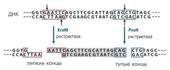
The end sequences will be the same when any DNA is cleaved with a given restriction enzyme and can be rejoined because they have complementary sequences. They can be ligated with DNA ligase to form a single molecule. Thus, it is possible to combine fragments of two different DNA and get the so-called recombinant DNA. This approach is used in the method of molecular cloning, which makes it possible to obtain individual genes and introduce them into cells that can form the protein encoded in the gene.
molecular cloning
Molecular cloning uses two DNA molecules - an insert containing the gene of interest, and vector- DNA acting as a carrier. The insert is "sewn" into the vector with the help of enzymes, obtaining a new, recombinant DNA molecule, then this molecule is introduced into host cells, and these cells form colonies on a nutrient medium. A colony is a progeny of one cell, i.e. a clone, all cells of the colony are genetically identical and contain the same recombinant DNA. Hence the term "molecular cloning", that is, obtaining a clone of cells containing a DNA fragment of interest to us. After the colonies containing the insert of interest to us are obtained, it is possible to characterize this insert by various methods, for example, to determine its exact sequence. The cells can also produce the protein encoded by the insert if it contains a functional gene.
When a recombinant molecule is introduced into cells, the genetic transformation of these cells occurs. Transformation- the process of absorption by a cell of an organism of a free DNA molecule from the environment and its integration into the genome, which leads to the appearance in such a cell of new heritable traits for it, characteristic of the organism-donor of DNA. For example, if the inserted molecule contains a gene for resistance to the antibiotic ampicillin, then the transformed bacteria will grow in its presence. Before transformation, ampicillin caused their death, that is, a new sign appears in the transformed cells.
VECTORS
A vector must have a number of properties:
First, it is a relatively small DNA molecule to be easily manipulated.
Secondly, in order for DNA to be preserved and reproduced in a cell, it must contain a certain sequence that ensures its replication (the origin of replication, or origin of replication).
Thirdly, it must contain marker gene, which ensures the selection of only those cells into which the vector has entered. Usually these are antibiotic resistance genes - then in the presence of an antibiotic, all cells that do not contain the vector die.
Gene cloning is most often carried out in bacterial cells, as they are easy to cultivate and multiply rapidly. In a bacterial cell, there is usually one large circular DNA molecule, several million base pairs long, containing all the genes necessary for bacteria - the bacterial chromosome. In addition to it, in some bacteria there are small (several thousand base pairs) circular DNA, called plasmids(Fig. 2). They, like the main DNA, contain a nucleotide sequence that provides the ability of DNA to replicate (ori). Plasmids replicate independently of the main (chromosomal) DNA, therefore they are present in the cell in a large number of copies. Many of these plasmids carry antibiotic resistance genes, which makes it possible to distinguish cells carrying the plasmid from normal cells. More commonly, plasmids carrying two genes conferring resistance to two antibiotics, such as tetracycline and amycilin, are used. There are simple methods for isolating such plasmid DNA free from the DNA of the main chromosome of the bacterium.

THE SIGNIFICANCE OF TRANSGENESIS
The transfer of genes from one organism to another is called transgenesis, and such modified organisms - transgenic. The method of gene transfer into microbial cells is used to obtain recombinant protein preparations for medicine, in particular, human proteins that do not cause immune rejection - interferons, insulin and other protein hormones, cell growth factors, as well as proteins for the production of vaccines. In more complex cases, when protein modification is carried out correctly only in eukaryotic cells, transgenic cell cultures or transgenic animals are used, in particular, livestock (primarily goats), which secrete the necessary proteins into milk, or proteins are isolated from their blood. This is how antibodies, blood clotting factors and other proteins are obtained. The transgenesis method produces cultivated plants that are resistant to herbicides and pests and have other useful properties. Using transgenic microorganisms to purify wastewater and fight pollution, there are even transgenic microbes that can break down oil. In addition, transgenic technologies are indispensable in scientific research - the development of biology today is unthinkable without the routine use of gene modification and transfer methods.
molecular cloning technology
inserts
To obtain an individual gene from any organism, all chromosomal DNA is isolated from it and cleaved with one or two restriction enzymes. Enzymes are selected so that they do not cut the gene of interest to us, but make breaks along its edges, and in plasmid DNA make one break in one of the resistance genes, for example, to ampicillin.
The molecular cloning process includes the following steps:
Cut and stitch - construction of a single recombinant molecule from an insert and a vector.
Transformation is the introduction of a recombinant molecule into cells.
Selection - selection of cells that received a vector with an insert.
cutting and stitching
Plasmid DNA is treated with the same restriction enzymes, and it turns into a linear molecule if such a restriction enzyme is selected that introduces 1 break into the plasmid. As a result, the same sticky ends appear at the ends of all the resulting DNA fragments. As the temperature is lowered, these ends join randomly and are ligated with DNA ligase (see Fig. 3).

A mixture of circular DNAs of different composition is obtained: some of them will contain a certain DNA sequence of chromosomal DNA connected to bacterial DNA, others will contain fragments of chromosomal DNA joined together, and still others will contain a reduced circular plasmid or its dimer (Fig. 4).

transformation
Next, this mixture is carried out genetic transformation bacteria that do not contain plasmids. Transformation- the process of absorption by a cell of an organism of a free DNA molecule from the environment and its integration into the genome, which leads to the appearance in such a cell of new heritable traits for it, characteristic of the organism-donor of DNA. Only one plasmid can enter and multiply in each cell. Such cells are placed on a solid nutrient medium containing the antibiotic tetracycline. Cells that did not get the plasmid will not grow on this medium, and the cells carrying the plasmid form colonies, each of which contains the descendants of only one cell, i.e. all cells in a colony carry the same plasmid (see Fig. 5).

Selection
Next, the task is to isolate only the cells into which the vector with the insert has entered, and to distinguish them from cells carrying only the vector without the insert or not carrying the vector at all. This process of selecting the right cells is called selection. For this, apply selective markers- usually antibiotic resistance genes in the vector, and selective media containing antibiotics or other selective substances.
In the example we are considering, cells from colonies grown in the presence of ampicillin are subcultured on two media: the first contains ampicillin, and the second contains tetracycline. Colonies containing only the plasmid will grow on both media, while colonies containing inserted chromosomal DNA in the plasmids will not grow on the medium with tetracycline (Fig. 5). Among them, those that contain the gene of interest to us are selected by special methods, grown in sufficient quantities, and plasmid DNA is isolated. From it, using the same restrictases that were used to obtain recombinant DNA, the individual gene of interest is cut out. The DNA of this gene can be used to determine the sequence of nucleotides, introduce into any organism to obtain new properties or synthesize the desired protein. This method of gene isolation is called molecular cloning.
FLUORESCENT PROTEINS
It is very convenient to use fluorescent proteins as marker genes in studies of eukaryotic organisms. The gene for the first fluorescent protein, green fluorescent protein (GFP) was isolated from the jellyfish Aqeuorea victoria and introduced into various model organisms (see Fig. 6) In 2008, O. Shimomura, M. Chalfi and R. Tsien received the Nobel Prize for the discovery and application of this protein.

Then the genes for other fluorescent proteins - red, blue, yellow - were isolated. These genes have been artificially modified to produce proteins with the desired properties. The diversity of fluorescent proteins is shown in fig. 7, which shows a petri dish with bacteria containing genes for various fluorescent proteins.

application of fluorescent proteins
The fluorescent protein gene can be fused with the gene of any other protein, then during translation a single protein will be formed - a translational fusion protein, or fusion(fusion protein), which fluoresces. Thus, it is possible to study, for example, the localization (location) of any proteins of interest in the cell, their movement. Using the expression of fluorescent proteins only in certain types of cells, it is possible to mark cells of these types in a multicellular organism (see Fig. 8 - mouse brain, in which individual neurons have different colors due to a certain combination of fluorescent protein genes). Fluorescent proteins are an indispensable tool in modern molecular biology.

PCR
Another method for obtaining genes is called polymerase chain reaction (PCR). It is based on the ability of DNA polymerases to complete the second strand of DNA along the complementary strand, as occurs in cells during DNA replication.
The origins of replication in this method are given by two small pieces of DNA called seeds, or primers. These primers are complementary to the ends of the gene of interest on two strands of DNA. First, the chromosomal DNA from which the gene is to be isolated is mixed with seeds and heated to 99 ° C. This leads to the breaking of hydrogen bonds and the divergence of DNA strands. After that, the temperature is lowered to 50-70 about C (depending on the length and sequence of seeds). Under these conditions, the primers are attached to complementary regions of chromosomal DNA, forming a regular double helix (see Fig. 9). After that, a mixture of all four nucleotides needed for DNA synthesis and DNA polymerase are added. The enzyme elongates the primers by building double-stranded DNA from the point of attachment of the primers, i.e. from the ends of a gene to the end of a single-stranded chromosome molecule. 
If the mixture is now heated again, the chromosomal and newly synthesized chains will disperse. After cooling, seeds will again join them, which are taken in large excess (see Fig. 10).

On the newly synthesized chains, they will join not to the end from which the first synthesis began, but to the opposite one, since the DNA chains are antiparallel. Therefore, in the second cycle of synthesis, only the sequence corresponding to the gene will be completed on such chains (see Fig. 11).

This method uses DNA polymerase from thermophilic bacteria that can withstand boiling and operates at temperatures of 70-80 ° C, it does not need to be added every time, but it is enough to add it at the beginning of the experiment. By repeating the heating and cooling procedures in the same sequence, we can double the number of sequences in each cycle, bounded at both ends by the introduced seeds (see Fig. 12).

After about 25 such cycles, the number of copies of the gene will increase by more than a million times. Such quantities can be easily separated from the chromosomal DNA introduced into the test tube and used for various purposes.
DNA sequencing
Another important achievement is the development of methods for determining the sequence of nucleotides in DNA - DNA sequencing(from English sequence - sequence). To do this, it is necessary to obtain genes pure from other DNA using one of the described methods. Then the DNA chains are separated by heating and a primer labeled with radioactive phosphorus or a fluorescent label is added to them. Please note that one seed is taken, complementary to one chain. Then DNA polymerase and a mixture of 4 nucleotides are added. Such a mixture is divided into 4 parts and one of the nucleotides is added to each, modified so that it does not contain a hydroxyl group on the third atom of deoxyribose. If such a nucleotide is included in the synthesized DNA chain, then its elongation will not be able to continue, because polymerase will have nowhere to attach the next nucleotide. Therefore, DNA synthesis after the inclusion of such a nucleotide is interrupted. These nucleotides, called dideoxynucleotides, are added much less than usual, so chain termination occurs only occasionally and in each chain in different places. The result is a mixture of chains of different lengths, each with the same nucleotide at the end. Thus, the chain length corresponds to the nucleotide number in the studied sequence, for example, if we had an adenyl dideoxynucleotide, and the resulting chains were 2, 7 and 12 nucleotides long, then adenine was in the second, seventh and twelfth positions in the gene. The resulting mixture of chains can be easily separated by size using electrophoresis, and the synthesized chains can be identified by radioactivity on X-ray film (see Fig. 10).

It turns out the picture shown at the bottom of the picture, called radioautograph. Moving along it from bottom to top and reading the letter above the columns of each zone, we will get the nucleotide sequence shown in the figure to the right of the autograph. It turned out that synthesis is stopped not only by dideoxynucleotides, but also by nucleotides in which some chemical group, for example, a fluorescent dye, is attached to the third position of the sugar. If each nucleotide is labeled with its own dye, then the zones obtained by separating the synthesized chains will glow with a different light. This makes it possible to carry out the reaction in one test tube simultaneously for all nucleotides and, by separating the resulting chains by length, to identify the nucleotides by color (see Fig. 11).

Such methods made it possible to determine the sequences not only of individual genes, but also to read entire genomes. Even faster methods for determining nucleotide sequences in genes have now been developed. If the first human genome was deciphered by a large international consortium using the first given method in 12 years, the second, using the second, in three years, now this can be done in a month. This allows you to predict a person's predisposition to many diseases and take measures in advance to avoid them.
31.2
For friends!
Reference
Molecular biology grew out of biochemistry in April 1953. Its appearance is associated with the names of James Watson and Francis Crick, who discovered the structure of the DNA molecule. The discovery was made possible through the study of genetics, bacteria and the biochemistry of viruses. The profession of a molecular biologist is not widespread, but today its role in modern society is very large. A large number of diseases, including those manifested at the genetic level, require scientists to find solutions to this problem.
Description of activity
Viruses and bacteria are constantly mutating, which means that medicines no longer help a person and diseases become intractable. The task of molecular biology is to get ahead of this process and develop a new cure for diseases. Scientists work according to a well-established scheme: blocking the cause of the disease, eliminating the mechanisms of heredity and thereby alleviating the patient's condition. There are a number of centers, clinics and hospitals around the world where molecular biologists are developing new treatments to help patients.
Job responsibilities
The responsibilities of a molecular biologist include the study of processes inside the cell (for example, changes in DNA during the development of tumors). Also, experts study the features of DNA, their effect on the whole organism and a single cell. Such studies are carried out, for example, on the basis of PCR (polymerase chain reaction), which allows you to analyze the body for infections, hereditary diseases and determine biological relationship.
Features of career growth
The profession of a molecular biologist is quite promising in its field and already today claims to be the first in the ranking of medical professions of the future. By the way, a molecular biologist does not have to stay in this field all the time. If there is a desire to change occupation, he can retrain as a sales manager for laboratory equipment, start developing instruments for various studies, or open his own business.